Phase differential microscopy is a technique employed to capture stunning images of crystals by leveraging the principles of phase contrast. When light passes through transparent crystals, variations in their refractive indices cause differences in the phase of the light waves. Specialized optical components, like phase plates or prisms, are utilized to manipulate these phase differences. As light interacts with the crystal, the phase-shifted waves create variations in brightness or contrast in the final image, enhancing the visibility of intricate crystal structures. This method enables photographers to capture detailed and vivid images of crystals without the need for staining or invasive techniques, providing a window into their natural beauty and complexity.
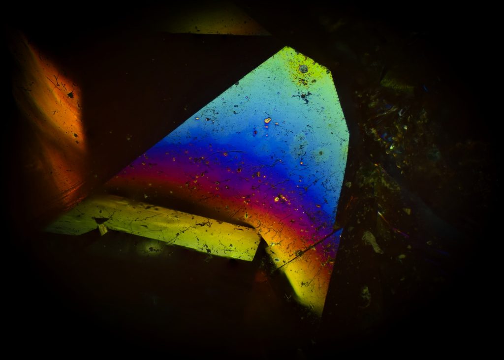
Here is a 96 image focus stack of a phase differential image I took of a crystal with my microscope, the color denotes depth. This is close to the limits of optical microscopy! Ultra high res version here:
High res: https://www.djdlabs.com/CRYSTAL.jpg
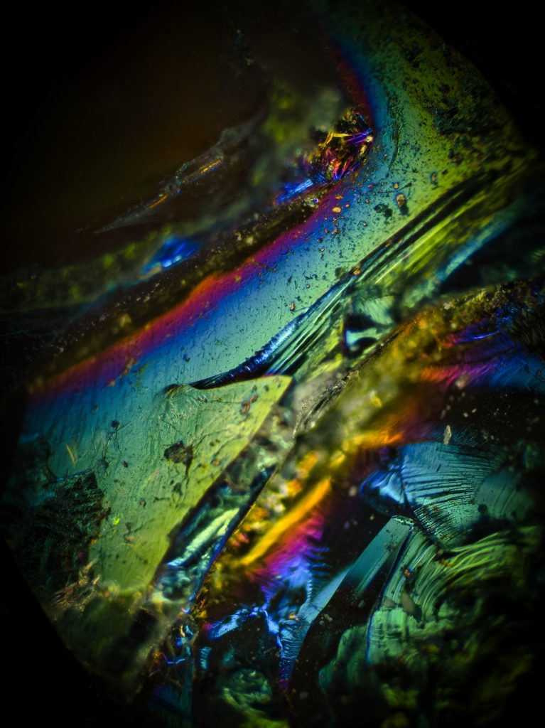
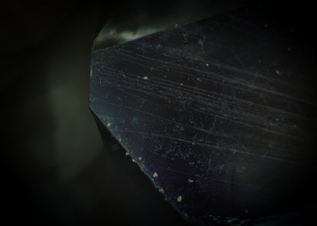
This last photo is the same crystal, but with visible light only! how cool is that!
These photos where taken with my Zeiss Aziomat, more about that here: https://www.djdlabs.com/zeiss-axiomat/

