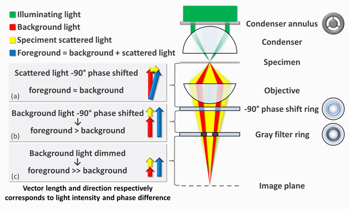
Phase-contrast microscopy is a powerful technique used to visualize unstained transparent specimens, such as biological cells or thin materials, by enhancing the contrast between different parts of the sample. In the context of visualizing stresses in a material, phase-contrast microscopy can be particularly insightful. When a material is subjected to stress, subtle changes occur in its refractive index due to variations in its physical properties, such as density or composition. Phase-contrast microscopy leverages these differences in refractive index to enhance the visibility of stress-induced alterations within the material. By carefully adjusting the optical components of the microscope, such as phase plates or annular diaphragms, phase shifts caused by stress-induced changes in refractive index can be converted into changes in brightness or contrast in the image. This allows researchers to observe and analyze stress patterns within the material, providing valuable insights into its mechanical properties and behavior under load. Overall, phase-contrast microscopy offers a non-destructive and highly sensitive method for visualizing and studying stress distributions in materials, contributing to advancements in materials science, engineering, and structural analysis.
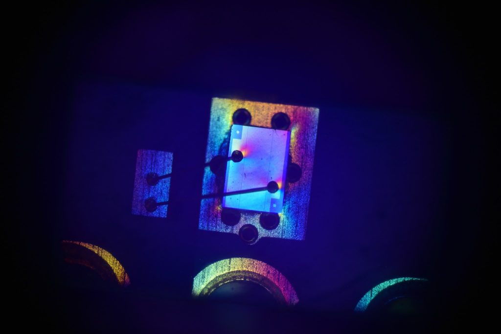
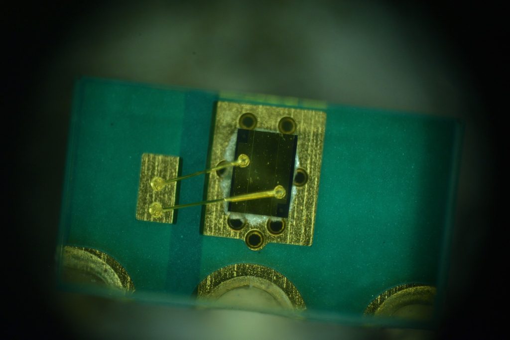
Here you can see the visible light and Phase Contrast photo of the same Laser Diode module under my Zeiss microscope, look close at where the wire bonds to the chips surface, you can see the stresses on the surface from the bonding wires with the exaggerated color change from the phase contrasted light.
External linked photo showing how the process works.
