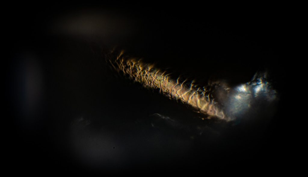
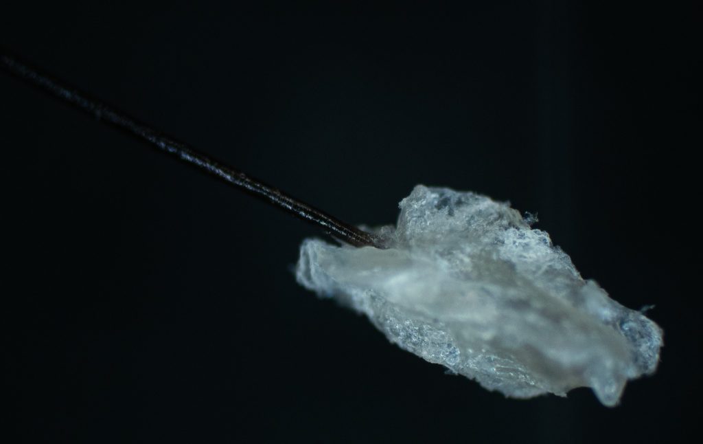
Capturing an incredible photo of a human hair under a microscope with the hair follicle intact was a moment of awe-inspiring precision. Using a high-resolution microscope equipped with a digital camera, I carefully positioned the hair sample and adjusted the focus to capture the intricate details of the hair shaft and follicle. The magnification revealed the remarkable complexity and structure of the hair follicle, highlighting its delicate features with stunning clarity. Remarkably, the human hair, with a diameter typically ranging from 0.02 to 0.18 millimeters, appears massive under the microscope’s lens, showcasing the remarkable capabilities of modern imaging technology to unveil the hidden wonders of the microscopic world. This photograph serves as a testament to the beauty and intricacy found within even the smallest components of the human body.


Observing the segmented lines on the hair under the microscope provides a fascinating glimpse into the intricate process of hair growth. These lines, known as “cuticle scales,” are the outermost layer of the hair shaft and play a crucial role in protecting the hair from damage and environmental stressors. Each cuticle scale overlaps with neighboring scales, forming a pattern akin to shingles on a roof. This arrangement helps to seal in moisture and prevent the hair from becoming brittle or frayed. Additionally, the direction and alignment of the cuticle scales can offer insights into the direction of hair growth and the overall health of the hair follicle. By closely examining these segmented lines, scientists and researchers can gain valuable information about the condition of the hair, including its strength, texture, and potential damage. Ultimately, studying the segmented lines on the hair provides a deeper understanding of the intricate biology behind hair growth and maintenance, shedding light on the factors that contribute to healthy, resilient hair.


